AIIMS-R Consultant Managed Life-Threatening Injuries by Self-Made firearm
Raipur
A 19-year-old engineering student who suffered injuries due to a self-made firearm was finally discharged by the All India Institute of Medical Sciences after 35 days of rigorous efforts in collaboration with different departments led by the surgery team. He is now out of danger with minimal risk of complications. The patient is on a feeding tube, and consultants are now planning to restore oral feed in the next few days.
The 19-year-old male engineering student was brought to the emergency department of AIIMS, Raipur, in a case of alleged self-inflicted firearm injury with a self-made weapon (a combination of high-impact penetrating and explosive devices) to the left upper abdomen on January 30, 2024.
At the time of presentation, the patient was alert and conscious, but his heart rate was in an increasing trend even after all management efforts, with gradual distension of the abdomen. After getting a call from Dr. Shivraj of Emergency, the operating surgeons, led by Dr. Radhakrishna Ramchandani, Associate Professor, Surgery Dept., along with Dr. Krishna Jangid, Dr. Tokho, and Dr. Aisharya, rushed into emergency and took the decision to operate the case immediately.
There were two wounds, one in the left middle abdomen (2 x 2 cm) with active oozing of blood and another (1 x 1 cm) just below the costal cartilage with tattooing of the left anterior abdominal wall. The exit wound was at the back, lateral to the spine. After seeing the device by the operating surgeon without delay, the decision for surgery, which is exploratory laparotomy, was made.
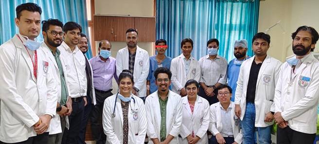
The surgery was performed by Dr. Ramchandani along with Dr. Jeevan Verma, Dr. Vishal Mohanty, Dr. Teja, and nursing officers Ms. Abhi and Ms. Moti. Anaesthesia was given by Dr. Mussavir and Dr. Prashant under the guidance of Dr. Chandan Dey, Assistant Professor of Anaesthesiology in Trauma and Emergency.
It had been anticipated about a major internal injury and sudden collapse of the patient due to a huge loss of blood immediately after laparotomy after knowing the mode of injury and after seeing the weapon by the operating surgeon. The team had been prepared accordingly. The things happen as per the team’s apprehension that there were around 2 litters of total blood loss, including blood clots, within 3 to 4 minutes before catching one injured major blood vessel.
This is often a huge challenge for any trauma surgeon to catch the major gush of bleeding points within minutes when you open the abdomen, which is already full of blood due to the sudden release of the tamponade effect after opening the abdomen. Surgeons managed to catch the active bleed within minutes, and blood pressure stabilized, which at the time was as low as 40/18 mm Hg.
The next step was to look at which vital organs were damaged and the extent of the damage. The surgery was most challenging as far as preoperative and postoperative mortality and morbidity were concerned. The exit wound was in the left paraspinal area, just touching the wall of the abdominal aorta. The patient was fortunate enough that if the abdominal aorta had been injured, he could not have reached the hospital.
The immediate life-threatening condition was managed. Both walls of the stomach were repaired. The near-complete transaction of duodenum was repaired. The management of its complications subsequently was a big challenge for the operating team to prevent secondary mortality and morbidity. In view of the site of injury and the type and mode of injury, multiple tubes through the nasal, oral, and abdominal were given.
Though the patient became hemodynamically stable after operation with support, he was kept in the ICU for 5 days in closed monitoring under the guidance of Dr. Chinmay Panda, Assistant Professor, Department of Anaesthesiology; Dr. Devendra; Dr. Balji; Dr. Sneha; and Dr. Hrishikesh; Dr. Abhinav; and Dr. Faizal, who were involved in post-operative management.
The next challenge was duodenal blowout, which is a sudden rupture of duodenum which is often life-threatening due to the sudden release of pancreatic juice and biles and all of its enzymes contained in this part of the small intestine. These could have been prevented because of the multiple tubes put inside the intestine and abdomen, which were helpful for diversion of juices and acids, decreasing the pressure of duodenum and maintaining nutrition as well. Again, early repair of duodenum within 24 hours, as in this case, decreases secondary mortality to 11% from 40%.
The next challenge was comorbidities due to this type of duodenal injury, which often causes a lot of complications like abdominal abscess, duodenal fistula, obstruction, and sepsis due to leakage. The literature says that in more than 66% of such cases, complications occur. But in this case, that could have been prevented by making the right decision about what to give and how to maintain each tube inserted inside the intestine and abdomen.
The crucial step of removing the duodenal tube was without any complications. The patient was discharged on day 35, when he was almost out of danger with minimal risk of complications.
According to Dr. Ramchandani, the next plan is to remove the feeding tube inside the intestine inserted through the abdominal wall only after a complete restoration of oral feed in the next few days. ‘This is one of the unique cases where the patient not only could have been saved, but he also recovered without any complications or comorbidities, he said. Lt Gen Ashok Jindal, Executive Director, congratulated the team for their collaborative efforts to save a precious engineering student’s life.